How should dogs (and other pets) be carried in the car according to the DGT?
According to the DGT, when travelling with your pet in the car, you must place a separator to prevent your pet from distracting you and

The use of the microscope and veterinary cytology allows for quick and accurate analysis of cells, tissues and biological samplesIt is a key tool for the diagnosis of infections, inflammations, masses, dermatological problems and other disorders in dogs and cats.
At Benivet Benissa we perform cytological studies as part of the diagnostic process, which allows us to obtain immediate information and guide the necessary treatment or complementary tests depending on each clinical case.
Benivet Benissa - Av. del País Valencià, 10, 03720 Benissa -
M-F 9:30-18:30 - S 10:00-13:00
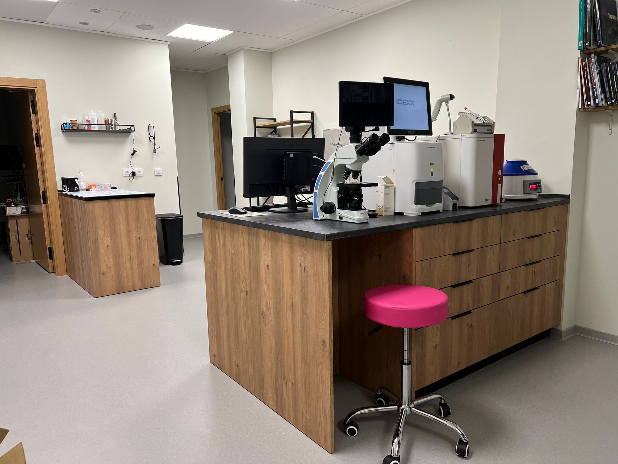
At Benivet Benissa we offer studies of veterinary cytology for dogs, cats and other pets, using the microscope as a key tool to detect infections, tumours and cellular alterations. This technique is minimally invasive, fast and very effective in obtaining crucial information about your companion's health.
Our team analyses samples of skin, lymph nodes, organs and body fluids with the support of image diagnosis and complementary tests of internal medicine. Thanks to this integration, we can provide accurate diagnoses and design the most appropriate treatment for each case.
Cytology is especially useful for lumps, non-healing wounds, inflammation or abnormal secretions. Early action can improve your pet's prognosis and quality of life.
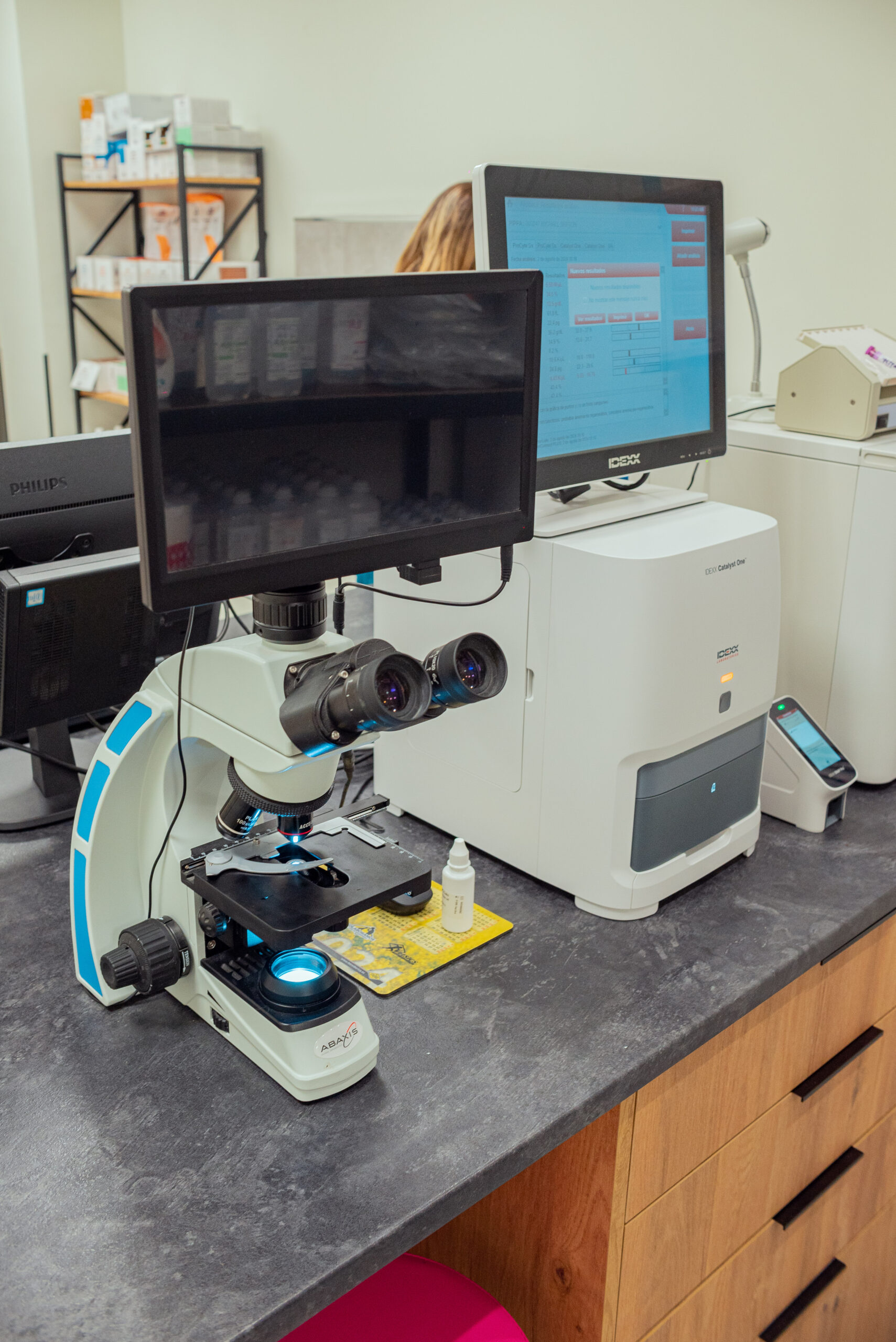
In our clinic, we use modern microscopy equipment to perform detailed and reliable cytological studies. This procedure, combined with services such as veterinary radiology or the ultrasoundhelps us to confirm diagnoses and plan effective treatments.
According to the Spanish Association of Small Animal Veterinary Specialists (AVEPA)cytology is a first-line diagnostic technique to evaluate lesions and masses in companion animals, as it provides rapid results and reduces the need for invasive procedures.
At Benivet BenissaAt our veterinary clinic, each pet receives a personalised approach, guaranteeing a fast, accurate diagnosis tailored to their needs. Your companion will be in the hands of a team committed to advanced veterinary medicine and animal welfare.
Veterinary cytology allows rapid cell analysis to guide diagnosis and clinical decision-making.
The use of the microscope allows diagnostic information to be obtained at the time of consultation, without the need to wait for external results.
Bacterial or fungal infections
Inflammatory processes
Initial diagnostic guidance
Cytology is particularly useful for studying lumps, nodules or visible lesions and deciding on the next steps.
Cutaneous or subcutaneous masses
Suspicious lesions
Differentiation between inflammatory and tumour processes
Cytology supports the work of other clinical areas and helps to adjust treatments more precisely.
Veterinary dermatology
Internal medicine
Treatment monitoring
Cytology and biopsy enable the examination of lesions, lumps and tissue abnormalities, but they do not analyse exactly the same thing.
The veterinary cytology examines cells obtained by fine-needle aspiration, a swab, a scraping or an impression. The biopsy a small sample of tissue is taken to analyse both the cells and the way in which they are organised.
The choice depends on the type of injury, its location and the information we need to obtain.
A smear test can help to identify:
Inflammatory processes.
Bacterial or fungal infections.
Yeasts and some parasites.
Accumulations of fat or cystic material.
Cellular changes associated with certain tumours.
Abnormalities in lymph nodes, organs or bodily fluids.
The type of cells present in a tumour.
In many cases, a smear test helps to guide the diagnosis and determine whether treatment should be started, a culture taken, the lesion monitored or further tests requested.
When the problem affects the skin, ears or hair, a cytology test may form part of the investigation carried out in veterinary dermatology.
If a lump is being investigated or a tumour is suspected, the case can be referred to veterinary oncology.
The technique depends on the injury being studied.
It can be used:
Fine-needle aspiration: to obtain cells from a lump, a lymph node or an organ.
Hyssop: commonly found in the ears, wounds, secretions and certain mucous membranes.
Scraping: used to treat certain skin conditions.
Direct printing: by placing a microscope slide over a superficial lesion.
Liquid samples: such as fluids found in cavities, joints or cystic lesions.
Many superficial samples can be taken during the consultation. In the case of internal lesions or those that are difficult to access, sampling can be carried out under guidance using veterinary ultrasound.
Ultrasound helps to locate the area and guide the needle towards the lesion, although the advisability of taking a sample must be assessed in light of its location, the patient’s condition and the potential risk of bleeding.
Not always. Some lesions contain cells with characteristics that are clear enough to guide or confirm the diagnosis, whilst others require a biopsy and histopathological examination.
Cytology mainly examines the appearance of cells. However, for some tumours, it is also necessary to analyse:
The structure of the fabric.
The relationship between cells and neighbouring structures.
The invasion of the margins.
The growth pattern.
The tumour grade, where applicable.
For this reason, a smear test may yield results such as:
Consistent with an inflammatory process.
Consistent with an infection.
Consistent with a benign lesion.
Suspected neoplasm.
Suitable for a specific type of tumour.
Inconclusive or non-diagnostic.
A ‘suspicious’ or ‘inconclusive’ result does not, on its own, confirm or rule out a tumour.
A sample may not provide sufficient information when:
It contains few cells.
It consists mainly of blood.
The lesion releases few cells.
The needle has not reached the target area.
There is inflammation or an infection masking the underlying condition.
The sample has deteriorated during preparation.
The cells in the lesion are difficult to distinguish.
It is necessary to examine the tissue’s overall structure.
In such situations, the team may recommend repeating the sample collection, using a different technique or sending the sample to a pathology laboratory.
The fact that the result is inconclusive does not necessarily mean that the injury is serious. It means that the sample obtained does not allow the clinical question to be answered with sufficient certainty.
A biopsy may be recommended when:
The smear test does not provide enough cells.
The result does not allow us to distinguish between various diseases.
The type of tumour needs to be confirmed.
It is important to study the architecture of the tissue.
The lesion grows in size or changes in appearance.
There is a wound that won’t heal.
The result will influence the planning of an operation.
It is necessary to analyse the margins after removing a batch.
A biopsy can be obtained by taking a small sample of the lesion or by removing it completely, depending on its size, location and the planned treatment.
If it is necessary to remove a mass or take a surgical sample, the case can be coordinated with veterinary surgery or with the Soft Tissue Surgery Unit.
When the procedure requires sedation or anaesthesia, the protocol is tailored to the patient by the department of veterinary anaesthesia and analgesia.
Yes. A smear test analyses the cells collected, but it does not always reveal the full extent of a lesion, its depth or its relationship to other organs.
Depending on the circumstances, the following may be recommended:
A veterinary ultrasound to examine organs, lymph nodes, bodily fluids and internal masses.
A veterinary X-ray to assess the chest, abdomen, bones or other structures.
A Veterinary CAT scan to delineate complex lesions and plan for sampling or a procedure.
Imaging tests can indicate where the abnormality is located and how extensive it is, whilst a smear test or biopsy helps to determine what type of cells or tissue it consists of.
A cytology test may reveal bacteria, fungi, yeasts and inflammatory cells, but it does not always make it possible to determine which microorganism is causing the problem or which medicine might be most suitable.
For certain infections, a culture may be recommended and, where appropriate, an antibiotic susceptibility test.
This crop can be particularly useful in the following situations:
Recurrent infections.
Wounds that do not respond to treatment.
Persistent ear infections.
Deep-seated infections.
Previous courses of antibiotics.
Suspected bacterial resistance.
The decision to carry out a culture depends on the microscopic findings, the progression of the condition and any previous treatments.
Once we have examined the sample, we will explain:
Which cells or microorganisms have been observed?
Whether the result helps to guide or confirm the diagnosis.
If the sample needs to be sent to a pathologist.
If necessary, repeat it.
Whether a biopsy, culture or imaging scan is required.
What treatment might be considered?
What checks are required for the injury?
In the case of diseases affecting internal organs or presenting complex symptoms, the results may be interpreted in conjunction with veterinary internal medicinea blood tests or other clinical tests.
Seek veterinary advice if you notice a lump that:
Grow or change quickly.
It becomes hard, lumpy or painful.
It becomes ulcerated, bleeds or produces discharge.
It makes it difficult to move, breathe or eat.
It is accompanied by weight loss or fatigue.
It reappears after having been removed.
It is located in an area that the pet constantly licks or bites.
The appearance, size or rate of growth do not provide a definitive indication of what type of lesion it is. Nor is it advisable to wait simply because the lump is small or does not seem to be causing any discomfort.
If your pet has already been assessed, please bring:
The cytological or histopathological report.
Slides, if available.
Photographs of the injury.
Image analysis and testing.
Surgical reports.
Treatments administered.
Information on how the lump has developed.
Vets who need to refer a case can use the service provided by Case studies from Benivet Benissa.
Have you noticed a lump, a wound that won’t heal or a recurring discharge? Request a veterinary assessment and smear test or call +34 622 325 271.
Benivet Benissa
Av. del País Valencià, 10, 03720 Benissa, Alicante
Telephone: +34 622 325 271
Opening hours: Monday to Friday from 09:30 to 18:30; Saturdays from 10:00 to 13:00.
 Publicado en Google
 Оксана Карпенко6 Octubre 2026
Trustindex verifica que la fuente original de la reseña sea Google.
Estamos muy agradecidos con el equipo de Benivet Benissa por la atención y el cuidado que han tenido con nuestro perro. Llegamos a la clínica después de varias semanas de problemas en la piel y de haber probado diferentes tratamientos sin conseguir una mejoría real. Nuestro Yorkshire, que ya tiene 14 años, tenía heridas, zonas muy inflamadas y pérdida de pelo. En Benivet realizaron las pruebas necesarias y finalmente encontraron la causa: una demodicosis grave con una infección bacteriana secundaria. Nos explicaron todo con mucha claridad y empezamos un tratamiento específico. Aunque el tratamiento todavía no ha terminado, la mejoría ha sido enorme: las heridas han desaparecido, la piel está mucho mejor, las zonas oscuras de las patas están desapareciendo y el pelo ya está empezando a crecer de nuevo. En la última revisión todavía quedaban algunas bacterias, por lo que nos han prolongado el tratamiento una semana más. Queremos agradecer especialmente la profesionalidad, la atención y la paciencia que han tenido con nosotros y con nuestro pequeño. Después de todo lo que habíamos pasado, por fin sentimos que nuestro perro está en buenas manos. ❤️ Muchas gracias a todo el equipo de Benivet Benissa. 🐶❤️Publicado en Google Ian Hunt Solo9 Septiembre 2026Had a CT scan on Tues, operation on Wed and they saved my dog! All of the team were exceptional! Thank you All!!Publicado en Google
 Willem Pieffers8 Septiembre 2026Durante los últimos dos meses hemos vivido momentos muy difíciles y llenos de incertidumbre. Nuestro galgo macho de 5 años se rompió una pata y además tenía un tumor. Para resumir una historia bastante larga: afortunadamente resultó no ser cáncer, pero finalmente tuvieron que amputarle la pata y ha pasado por momentos de mucho dolor. Estamos enormemente impresionados y agradecidos con todo el personal de esta clínica. Tanto el equipo de veterinarios como el personal de apoyo se comunican perfectamente, son muy empáticos y atentos, pero al mismo tiempo proactivos y tremendamente profesionales. Sinceramente, no tenemos ni un solo punto de crítica. Estamos simplemente muy felices de haber llegado a esta clínica y, sin ninguna duda, a partir de ahora serán nuestros veterinarios de confianza. **Gracias por todo, querido equipo de la Clínica Veterinaria Benivet.** ❤️ Willem y JaviPublicado en Google
 Vanessa Torres Martínez8 Septiembre 2026Excepcional es poco. Fuimos por una urgencia y no pude quedar más encantada. Trato súper genial, seguimiento super y personalmente volvería 100%Publicado en Google
 Sixtine Lamarque15 Agosto 2026Fuimos el viernes por la tarde porque nuestro gato se comió un hilo largo. Las veterinarias fueron súper pacientes a pesar de que el gato se puso peleón por el miedo. Le hicieron ecografía y luego lo metieron a cirugía casi inmediatamente. El cirujano fue excelente, nos explicó todo el procedimiento y nos avisó por WhatsApp cuando estuvo listo el gatito, se quedó en observación y al día siguiente lo hemos recogido. La operación fue impecable y el gatito está en casa, la sutura está perfecta. Fueron excepcionales.Publicado en Google
 Alejandro Blázquez15 Agosto 2026Lleve a mi gato de 6 años de urgencia porque se había tragado un hilo largo y gordito, y por la mañana vomitó un trozo. Menos mal que lo hice ya que al día siguiente era festivo, y haber esperado más habría resultado seguramente en necrosis y perforaciones intestinales. Mi gatito fue muy bien atendido por todos los veterinarios. Lo sedaron porque estaba nervioso, le hicieron ecografía para encontrar el hilo en el yeyuno, ya medio plegado, y lo operaron. Fue caro pero lo pagaría de nuevo, porque la vida de mi gatito no tiene precio y lo atendieron aún cuando iban a empezar después de su hora de cierre. Además está mañana al recogerlo estaba bien, tranquilo y dolorido, y la sutura se ve super limpia y bien hecha. Mis agradecimientos a Constantino y al resto de veterinarias que atendieron a mi gato.Publicado en Google
 Roger Llorens Salvador14 Agosto 2026No tenemos más que agradecimiento para todo el equipo de Benivet Benissa. Llevamos a nuestro perro Dachs para una cirugía de columna delicada —una hernia que le comprimía la médula— y desde el primer minuto nos transmitieron una tranquilidad enorme. Incluso nos avisaron de la dirección exacta para no confundirnos con otra clínica que hay a pocos metros: un detalle pequeño que ya dice mucho de cómo trabajan. Queremos destacar especialmente a dos personas. Al traumólogo, Constantino, que hizo un trabajo impecable: retiró toda la hernia y liberó la médula sin complicaciones. Y a la anestesista, Ana, que cuidó cada detalle —algo nada menor en una intervención así—. Gracias a ambos, en las primeras 24 horas Dachs ya recuperaba movilidad voluntaria en la pata. Cuando surgió un pequeño imprevisto con la sonda, lo resolvieron al momento y con total profesionalidad. Pero si algo destaca por encima de lo médico es el trato humano de todo el equipo. Nos mantuvieron informados con mensajes constantes y cercanos, nos escribían para contarnos que Dachs "se portaba muy bien", que nos deseaba buenas noches, e incluso se adelantaron ofreciéndonos un arnés de apoyo por si lo necesitaba. Se nota que aquí no solo tratan a los animales: los cuidan de verdad. Gracias de corazón a Constantino, a Ana y a todo el equipo de Benivet.Publicado en Google
 Jorge Sausalima13 Agosto 2026Muy contento con benivet, un buen servicio y mis animales están en muy buenas manos con ellas. De lo mejor de la zona.
The microscopy and veterinary cytology allow us to obtain quick and very useful information on inflammation, infection and cell typesespecially in problems of skin, ear and lumps. At Benivet Benissa we indicate it after the examination when we need to guide the diagnosis and treatment. If it is necessary to complete the study, we can recommend the following tests only when they provide useful information:
Veterinary practice in BenissaComplete examination and decision of the most appropriate test according to the case (skin, otitis, lumps, non-healing wounds).
Veterinary dermatologywhen there is persistent itching, recurrent dermatitis, alopecia, bad odour, crusting, or repeated otitisfor a medium-term diagnosis and monitoring plan.
Blood testsuseful if the problem is recurrent, there are general signs (fever/decrease/weight loss), or we need to assess the safety of treatments.
Stool analysis/coprological testsIf there is recurrent diarrhoea or suspicion of parasites that may influence the picture (e.g. perianal itching or associated digestive problems).
Veterinary ultrasoundif there are internal masses, abdominal pain or we need to assess organs and soft tissues.
If the case requires a global approach or follow-up of a disease, we coordinate the plan from veterinary internal medicine. And if pain or inflammation is a condition for well-being, we can rely on the PAIN UNIT for safe pain management.
👉 If your pet has a lump, recurrent otitis or recurring skin problems, request an appointment here or call +34 622 325 271.
According to the DGT, when travelling with your pet in the car, you must place a separator to prevent your pet from distracting you and
Only a veterinarian can assess the health status of the animals, taking also into account other circumstances such as the type of
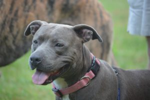
Domestic or companion animals, particularly those belonging to the canine species, shall also be classified as potentially dangerous,
A smear test helps guide the diagnosis, but does not always confirm it. Depending on the result, imaging tests, a biopsy or follow-up with a specialist may be recommended.
Cytology forms part of the investigation into itching, recurrent ear infections, alopecia and skin lesions.
When a lump is being investigated or a tumour is suspected, the case is managed by the oncology department.
It enables the localisation of internal masses and guides the needle insertion in hard-to-reach lesions.
If the problem is recurrent, there are general symptoms, or the safety of a treatment needs to be assessed.
When it is necessary to remove a mass or take a surgical biopsy in order to confirm the diagnosis.
Joint interpretation of results in cases of diseases affecting internal organs or involving complex symptoms.
Cytology is a diagnostic technique that analyses cells obtained by puncture, swabs or scrapings. It allows the identification of infections, inflammations, parasites, fungi and, in many cases, whether a lump is benign or suspicious for malignancy. It is a quick, painless and very useful test for initial diagnosis.
These are procedures minimally invasiveThe procedure is comparable to an injection. Sedation is not needed in most pets; only in very nervous animals or when the area is painful. The test is safe and generally well tolerated.
It is indicated in cases such as:
Lumps or masses in skin or subcutaneous tissue
Recurrent otitis
Non-healing wounds
Skin inflammations
Enlarged nodes
Suspected bacterial or fungal infections
Cytology helps to guide the diagnosis and to decide whether additional tests such as biopsy, culture or CT scan are needed.
The microscope makes it possible to assess real-time sampleswhich speeds up the diagnosis. We can see:
Inflammatory cells
Compatible tumour cells
Fungi and yeasts
Skin parasites
Bacteria
Sample quality and degree of infection
This makes it easier to tailor treatment from the first visit.
Cytology provides valuable information and, in many cases, allows differentiation between benign and malignant lesions. However, some tumours require biopsy and histopathological analysis to obtain a definitive diagnosis. At Benivet Benissa we will tell you when it is necessary to extend the study.
In most cytologies the preliminary result is obtained in the same consultationthanks to the immediate microscopic examination. In complex cases we send the sample to a specialised pathologist, who issues a detailed report. We explain the findings in a clear way and give you guidance on the next steps.
Do you have any doubts about care, treatment or prevention for your pet? Consult our section of Frequently Asked Questions where we solve the most common queries about dogs and cats.