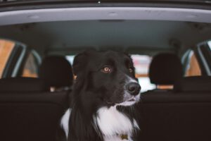
How should dogs (and other pets) be carried in the car according to the DGT?
According to the DGT, when travelling with your pet in the car, you must place a separator to prevent your pet from distracting you and

The veterinary radiology is an essential diagnostic imaging test to evaluate bones, joints, thorax and abdominal cavity in dogs and cats, allowing the rapid and accurate detection of fractures, bone alterations, respiratory problems and other internal pathologies.
At Benivet Benissa we carry out radiological studies as part of the diagnostic process, always integrated with the clinical examination and other complementary tests, in order to obtain a complete and reliable assessment of each patient.
Benivet Benissa - Av. del País Valencià, 10, 03720 Benissa -
M-F 9:30-18:30 - S 10:00-13:00
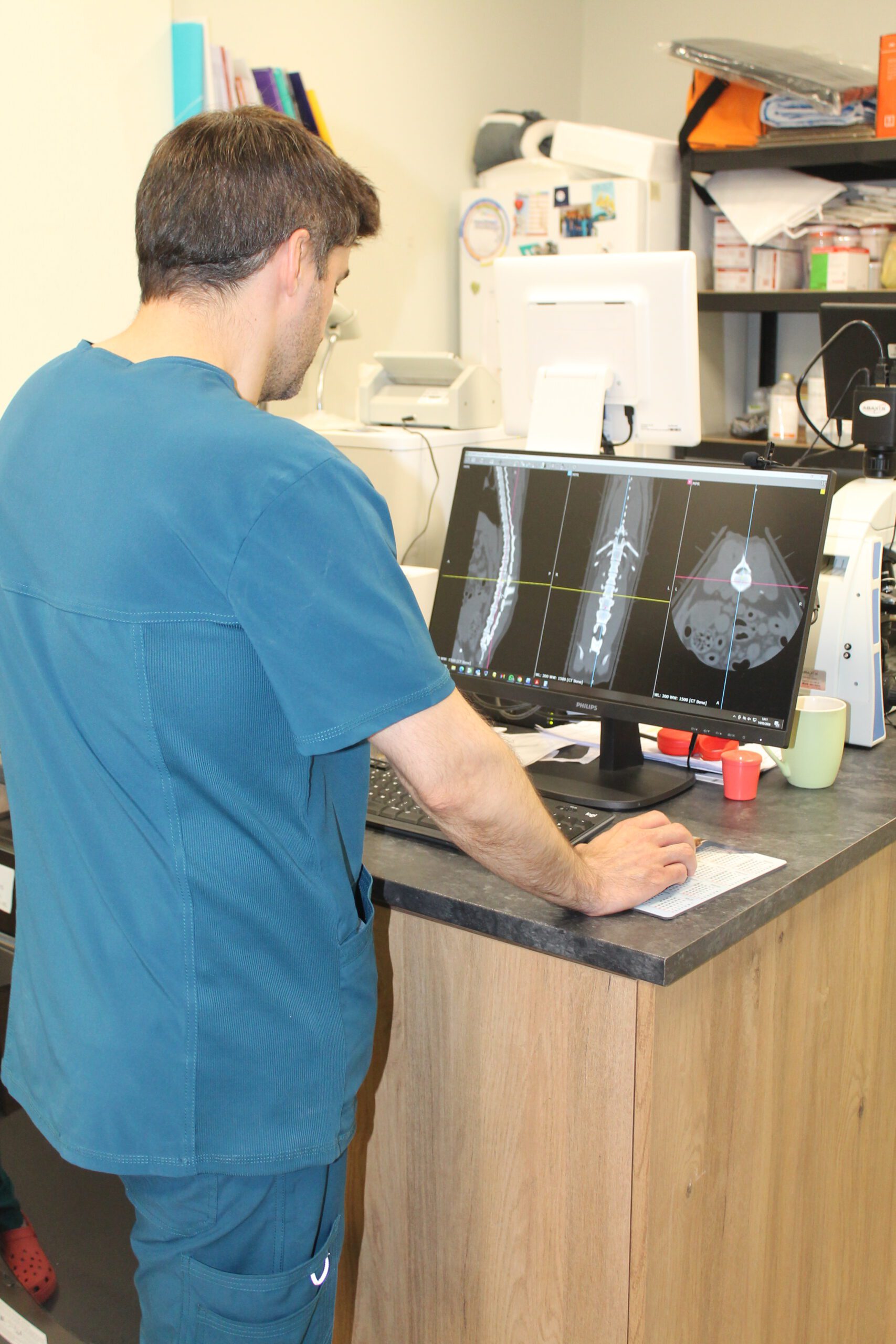
The veterinary radiology is an essential technique for diagnosing internal diseases and injuries in dogs, cats and other pets. Thanks to the ability to obtain accurate images of bones, joints and internal organs, veterinarians can detect problems that are not visible to the naked eye.
At Benivet BenissaWe have state-of-the-art equipment for digital radiographyThe results are clear and rapid. This facilitates the identification of fractures, respiratory pathologies, digestive disorders, joint diseases, dental problems or even internal tumours.
Radiology is an integral part of our image diagnosis and is usually complemented by the general medicineIt also plays a key role in the diagnosis and treatment of diseases, helping to make complete examinations and better clinical decisions. In addition, it plays a key role in veterinary dentistrywhere dental X-rays allow an in-depth evaluation of the bone structure.
At our veterinary clinic in Benissa, we perform X-rays with state-of-the-art digital equipment, designed to guarantee the safety of your pet and offer high resolution images with minimum exposure to radiation.
The process includes rapid preparation, proper positioning of the patient for optimal imaging and real-time analysis by our veterinary team. This allows us to provide reliable diagnoses in a matter of minutes and define the most appropriate treatment.
In cases that require it, we collaborate with imaging specialists to confirm findings and ensure maximum accuracy. Depending on the European College of Veterinary Diagnostic Imaging (ECVDI)Digital radiology is an essential tool in modern veterinary medicine for early diagnosis and monitoring of multiple pathologies.
At Benivet Benissaa quick and safe diagnosis makes all the difference in your pet's care.
Veterinary radiology is a quick and effective test to evaluate internal structures, especially bones, joints and thorax.
X-rays are essential to assess alterations of the skeletal system and to detect traumatic or degenerative lesions.
Fractures and dislocations
Sciatica and joint problems
Assessment of osteoarthritis or other bone pathologies
It allows to evaluate internal structures and to detect alterations in the thorax and abdomen quickly.
Pulmonary and cardiac study
Radiographically visible abdominal abnormalities
Clinical diagnostic support
Radiology is used as an adjunct to clinical examination and other diagnostic tests.
Confirmation or exclusion of diagnoses
Follow-up after treatment or surgery
Coordination with ultrasound or CT
An X-ray transforms a three-dimensional area of the body into a two-dimensional image. For this reason, bones, organs and tissues may appear superimposed on one another, and a single image does not always allow a lesion to be correctly located.
Producing several projections from different angles helps to provide a more comprehensive view and to interpret the findings more effectively.
When investigating a lameness, a fracture or a joint, it is often necessary to obtain images from more than one angle.
These projections can help to assess:
The location and orientation of a fracture.
The alignment of the bones.
The condition of a joint.
The presence of osteoarthritis, dislocations or bone abnormalities.
The stability of a surgical repair.
The progression of the injury during check-ups.
In some cases, it may also be useful to compare the affected limb with the opposite one.
The examination and interpretation of the images can be coordinated with the veterinary orthopaedics and traumatology.
If a complex lesion or one requiring surgical planning is detected, the case can be assessed from the Orthopaedics, Traumatology and Spinal Surgery Surgical Unit.
To examine the heart, lungs, trachea and other thoracic structures, a number of images may be required with the patient positioned in different ways.
This makes it possible to assess the following more accurately:
Changes in the lungs.
Heart size and shape.
Presence of fluid or air in the chest.
Masses or mediastinal abnormalities.
Injuries caused by trauma.
Progression of a respiratory or cardiac condition.
The position affects how air, blood and organs are distributed within the chest. For this reason, an abnormality may be more clearly visible in one projection than in another.
Where there is a cough, fatigue, shortness of breath or suspected heart disease, X-rays may be supplemented by an assessment of veterinary cardiology o internal medicine.
Abdominal X-rays can provide information on the size, position and shape of certain organs, as well as helping to detect specific foreign bodies, stones, gas accumulations or abnormalities in the digestive contents.
However, organs and soft tissues appear similar on a conventional X-ray. Therefore, depending on the findings, it may be advisable to supplement the examination with a veterinary ultrasound.
Combining these two techniques can help to assess:
Vomiting or abdominal pain.
Suspected foreign body.
Stomach or bowel distension.
Changes in organ size.
Urinary problems.
Presence of fluid or abdominal masses.
If there is difficulty urinating, blood in the urine or a suspected urinary stone, the examination can be arranged with veterinary urology and be complemented by a urinalysis.
For an X-ray to be useful, the area to be examined must be positioned in a specific way. Any rotation, movement or incorrect positioning may obscure a lesion or make it difficult to compare images.
During the test, the team tries to achieve the required position carefully and in the shortest possible time.
You should not force an animal when:
He is experiencing severe pain.
He may have a fracture.
He is experiencing breathing difficulties.
He’s either very scared or trying to escape.
He is unable to maintain the required posture.
Very accurate measurements or projections are required.
No. Many X-rays can be taken whilst the animal is awake, particularly when it is calm and the position does not cause it any discomfort.
Sedation may be considered when:
The movement prevents the capture of interpretable images.
Touching the area is painful.
Very precise positions are required.
Fear or anxiety increases the risk to the patient.
A longer-term study is needed.
This is an animal that does not tolerate being restrained.
The decision is made on a case-by-case basis. When sedation is required, the protocol is coordinated with the veterinary anaesthesia and analgesia.
Not necessarily. An image may be repeated because:
The patient has moved.
The section was not positioned correctly.
We need a different perspective.
A structure appears superimposed.
A specific section needs to be expanded.
We want to verify a questionable finding.
It is necessary to compare the results with those of a previous study.
The aim is to obtain images of sufficient quality to answer the clinical question that prompted the test.
No. An X-ray can confirm certain abnormalities, help guide the diagnosis or rule out some problems, but it cannot identify all diseases.
It may be necessary to supplement the study by:
Veterinary ultrasound to examine organs and soft tissues.
Veterinary CAT scan to examine the skull, spine, complex joints or specific masses in greater detail.
Blood tests to assess the general condition and functioning of various organs.
Veterinary cytology or a biopsy when it is necessary to determine the nature of a lesion.
Other tests selected on the basis of symptoms and the results obtained.
The images should be interpreted in conjunction with the examination, medical history and other clinical tests. The presence of a radiographic abnormality does not necessarily mean that the pet requires surgery.
If your dog or cat has been treated at another clinic, please bring:
The original X-ray images.
The interpretation report.
Previous analyses and tests.
Surgical reports.
Treatments administered.
Approximate date on which the symptoms first appeared.
Wherever possible, please provide the complete digital files rather than just screenshots. Comparing scans can help assess progress and avoid unnecessary repeat examinations.
Veterinary professionals can also submit the documentation via the Case studies from Benivet Benissa.
Does your pet need an X-ray, or would you like to review a previous X-ray? Book an appointment at Benivet Benissa or call +34 622 325 271.
Benivet Benissa
Av. del País Valencià, 10, 03720 Benissa, Alicante
Telephone: +34 622 325 271
Opening hours: Monday to Friday from 09:30 to 18:30; Saturdays from 10:00 to 13:00.
Trustindex verifica que la fuente original de la reseña sea Google. Muchas gracias a toda la gente que trabaja en Benivet ☺️ Son profesionales y gente muy amable. Mi gata Mini está mucho mejor gracias a ellos. Son un gran equipo y el trato a los animales no puede ser mejor, los tratáis genial. Sin duda siempre volvemos!!Publicado en GoogleTrustindex verifica que la fuente original de la reseña sea Google. Todo genial! Muy recomendable.Publicado en GoogleTrustindex verifica que la fuente original de la reseña sea Google. Súper professionals, es nota que els agrada el que fan. Molt contenta.Publicado en GoogleTrustindex verifica que la fuente original de la reseña sea Google. Atención inmejorable, profesionalidad indiscutible!! Son un equipo de 10! Conocí a Ana y Andrea hace ya 4 años y estoy encantada de poder contar con ellas y confiarles la salud de mis mascotas! Transmiten el amor por la profesion y los animales!! Es un placer poder contar con todo el equipo!! Muchas gracias chic@s !!Publicado en GoogleTrustindex verifica que la fuente original de la reseña sea Google. Onze hond had een splinter in zn poot, we konden direct terecht en uurtje later konden we hem weer ophalen, operatie en een narcoseprik samen voor maar €. 90.00 maar wat een lieve maar vooral professionals......TOPPublicado en GoogleTrustindex verifica que la fuente original de la reseña sea Google. Para mí es el mejor veterinario de la zona , siempre el mejor trato y con los mejores resultados .Publicado en GoogleTrustindex verifica que la fuente original de la reseña sea Google. This vet is amazing. We travel from Calpe to Benissa for their services. The staff are friendly, knowledgeable and so helpful.Publicado en GoogleTrustindex verifica que la fuente original de la reseña sea Google. Estoy muy contenta con este veterinario, la atención siempre ha sido muy buena y personalizada y las veterinarias tratan a Queen con mucho cariño, a pesar de sus ladridos :)Publicado en GoogleTrustindex verifica que la fuente original de la reseña sea Google. Super cercanos, dispuestos siempre a ayudarte y muy pendientes de los animalitos. Son un equipo increíble!
The veterinary radiology (X-rays) is a quick and very useful test to evaluate bone, joints and thoraxIt can also help with some abdominal problems (depending on the case). At Benivet Benissa we indicate it when, after the examination, we need to confirm a suspicion (trauma, lameness, cough, respiratory difficulty, pain, etc.). If it is necessary to complete the study, we can recommend the following tests only when they provide useful information:
Veterinary practice in BenissaComplete examination and indication of the most appropriate test according to symptoms.
Veterinary orthopaedics and traumatologywhen there is lameness, joint pain, muscle/bone injury or suspected osteoarthritisto assess the treatment and follow-up plan.
Blood testsuseful if there is fever/decompression, suspected systemic inflammation or to assess safety of medication and/or sedation if required.
Veterinary ultrasoundkey complement when the problem is in organs and soft tissues (abdomen, bladder, liver, kidneys) or when radiography and ultrasound are needed to complete the information.
Veterinary CAT scan: recommended in complex cases (spine, joints, skull) when we need more detail to decide on treatment or plan surgery.
If the case requires a global approach or chronic disease monitoring, we coordinate the plan from veterinary internal medicine. And if there's a procedure or intervention, we integrate it with anaesthesia and analgesia for safe pain management.
👉 If your pet has had a blow/fall, is limping, in pain, has a persistent cough or difficulty breathing, request an appointment here or call +34 622 325 271.
According to the DGT, when travelling with your pet in the car, you must place a separator to prevent your pet from distracting you and
Only a veterinarian can assess the health status of the animals, taking also into account other circumstances such as the type of
Domestic or companion animals, particularly those belonging to the canine species, shall also be classified as potentially dangerous,
Here is our timetable
An X-ray can confirm or rule out many conditions, but not all of them. Depending on the findings, the investigation is supplemented by further tests or consultations with other specialists.
A key diagnostic tool when the problem lies in organs and soft tissues, which appear similar on an X-ray.
When more detail is required for the skull, spine, complex joints or specific masses.
Interpretation of imaging findings relating to lameness, fractures and joints, and a treatment and follow-up plan.
Sedation when pain, fear or the need for specific positioning prevent useful images from being obtained.
Useful in cases of fever or malaise, suspected systemic inflammation, or to assess the safety of sedation.
In cases of cough, fatigue or suspected heart disease, chest X-rays are supplemented by a cardiological assessment.
Radiography allows the evaluation of structures such as bones, joints, thorax, lungs, trachea, abdomen and spine. It is a key tool in cases of lameness, fractures, respiratory problems, foreign body ingestion, abdominal masses or cardiac alterations evidenced by imaging.
Yes. Veterinary X-rays use very controlled doses X-rays and are performed according to strict safety protocols. Exposure is brief and limited to the necessary area. At Benivet Benissa we have direct digital radiologywhich reduces time and improves image quality.
Not always. Most pets can be x-rayed without sedation. However, if the animal is very nervous, the area is painful to manipulate, or very specific projections are needed, a light sedation to obtain quality images and avoid stress.
X-rays are usually obtained in few minutesand digital images can be viewed instantly. The vet usually discusses the findings during the same visit and, if necessary, suggests additional tests such as ultrasound or blood tests.
In many cases, yes. Radiography helps to identify masses, bony changes, pulmonary changes, foreign bodies, fluid in the chest or abdomenamong others. Depending on the type of injury, it may require a ultrasound or CT scan for further study.
Digital radiology offers higher resolutionThe results are quicker, with less exposure, the ability to adjust contrast and brightness without retakes, and faster results. This translates into a quicker and more accurate diagnosis for your pet.
Do you have any doubts about care, treatment or prevention for your pet? Consult our section of Frequently Asked Questions where we solve the most common queries about dogs and cats.